Better safe than sorry: the bizarre sex life of mayflies
12.03.2026 | M.Sc. Raffaele GambaMayflies are a symbol of transience. As adult insects, they often get only a single evening in which everything counts: finding a partner, mating, and laying eggs. Just how bizarre this can be becomes clear when you take a look at their genitalia—and at our CT data.

(Copyright: A. Staniczek / SMNS)
A life that builds up to its last day
A mayfly spends most of its life as a larva in freshwater; depending on the species, this can last from months to several years. The final larval stage is already fully geared toward the last stretch as a winged insect: feeding stops, females begin building up egg reserves, and the wings complete their growth.
Once it has molted into an adult (Fig. 1), there is only one task left: reproduction. Feeding is no longer possible—literally. The mouthparts are reduced, and the gut is sealed off at both ends. Instead, it is repurposed as an air-filled balloon that saves weight while simultaneously stiffening the abdomen. Now the clock is ticking …
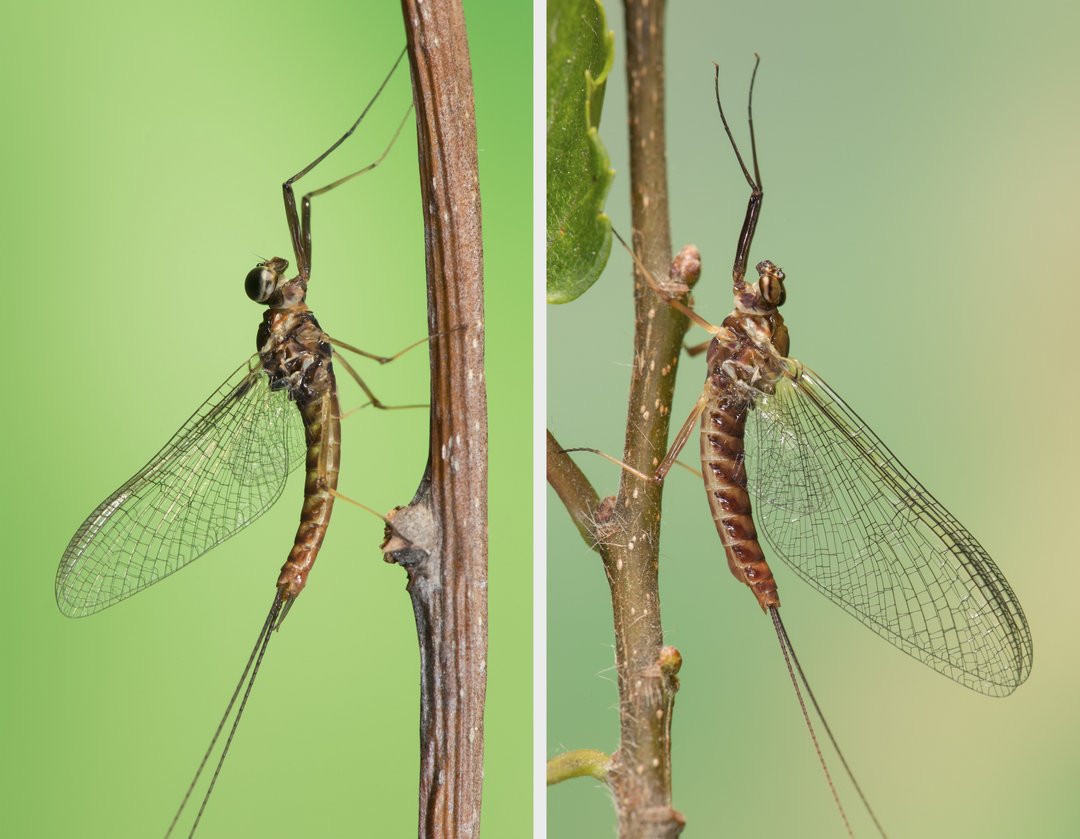
Figure 1: Male (left) and female (right) of the mayfly species Ecdyonurus venosus, body length approx. 1.2 cm. (Copyright: A. Staniczek / SMNS)
“Swarm Kamasutra”: an acrobatic aerial mating ritual
When conditions are right, often during calm evenings and preferably after a rain shower, males form swarms over bodies of water to attract females. As soon as a female enters the swarm, mating begins immediately, mid-air.
A male approaches the female from below and grasps her with forelegs elongated specifically for this purpose, gripping her at the wing bases. He then curves his abdomen forward and upward by almost 180° and secures his hold with specialized genital forceps (claspers) (Fig. 2). Acrobatically anchored to the female in this way, copulation can proceed.
Unlike more ancient insect lineages that deposit spermatophores which are then taken up by females, mayflies, as the evolutionarily oldest branch of flying insects, were effectively the first to evolve direct mating with internal fertilization. If you want to understand how complex genitalia and their functions evolved in flying insects, mayflies are a great place to start.

Figure 2: Ecdyonurus venosus mating. The female is on top, the male below. (Copyright: A. Staniczek / SMNS)
Insects in the CT scanner
Until now, almost nothing was known about mating in mayflies for a simple reason: copulation is brief, and it doesn’t happen in a convenient location, but high up in the air. So how do you get a copulating pair from mid-air into a CT scanner for analysis?
We tried our luck in the Black Forest, searching for Ecdyonurus venosus. To catch a copulating pair, we needed a net with a long handle—a 2.5-meter handle worked well. Even then, we often failed because the animals reacted to the disturbance in a decidedly unromantic fashion: most pairs separated immediately.
If they didn’t break off, we used a method borrowed from sports medicine: with freezing spray, we literally froze the pair mid-mating (Fig. 3). After shock-freezing, we carefully transferred them, still in copula, into a special fixative and ultimately stored them in ethanol.
To examine what happens inside the female during mating, we used synchrotron X-ray microtomography (µCT) at the synchrotron particle accelerator of the Karlsruhe Institute of Technology (KIT). The synchrotron produces very uniform, high-energy X-ray radiation, and the specimen is imaged while being rotated step by step. From over 1,000 individual images, a 3D model can then be reconstructed on the computer. In principle, it’s the same as a CT scan in a hospital, just at a much higher resolution, revealing structures down to the micrometer scale. The advantage: you can dissect the data digitally and create 3D models without destroying the fragile animals.

Figure 3: Benedict Stocker shock-freezing a copulating pair of mayflies in a net. (Copyrigth: A. Staniczek / SMNS)
Genital forceps and a paired penis with extendable sex spines
Males are equipped with a paired penis on the underside of the ninth abdominal segment: two separate penis lobes, each with its own genital opening, arise from a common shaft. Between them two spines, only their tips visible at rest, are located whose role in mating was unknown until now. On either side of the paired penis are forceps-like claspers fused at the base (Fig. 4). With these structures, the male can hold onto the female during mating.
In females, externally, there’s little to see apart from the membranous, rough subgenital plate (Fig. 5). This covers a copulatory pouch that opens to the rear, and the paired oviducts open into it separately.

Figure 4: Ventral view of the end of the male abdomen with paired reproductive organ and long claspers, SEM (scanning electron microscope). (Copyright: R. Gamba / SMNS)

Figure 5: Ventral view of the end of the female abdomen with transversely furrowed subgenital plate, SEM. (Copyright: R. Gamba / SMNS)
Anchor, stretch, fertilize
µCT scans show that the penis changes shape during mating (Fig. 6): powerful muscles cause a deformation of the penis shaft, making the penis lobes fold over. At the same time, the penial spines extend and prick into the thin membrane of the female’s copulatory pouch (Fig. 7). This stretches the pouch so that it can receive large amounts of sperm, which are stored in a folded membrane at the front of the copulatory pouch.
The penial spines also ensure that the organ is securely anchored during copulation, keeping it from slipping out during flight maneuvers. Since other males often try to disrupt the pair and steal the female, secure anchorage pays off even more.

Figure 6: Penis of Ecdyonurus venosus, lateral view. Left: at rest; right: during mating with extended penial spines. Reconstruction from µCT data; penis length approx. 0.8 mm. (Copyright: B. Stocker / R. Gamba / SMNS)

Figure 7: Penial spines of the mayfly Ecdyonurus venosus during mating. µCT image, looking rearward from inside the female. (Copyright: T. van de Kamp / KIT)
Graphical overview of the mechanisms underlying penile shape change. Left: µ-CT reconstruction of the penis in its resting configuration; right: µ-CT reconstruction of the penis in its active configuration. In between is explanatory text: The penes of Ecdyonurus spp. transition into a copulatory configuration during mating. 1: The intrinsic penis muscle contracts, the penis shaft is deformed 2: The penis lobes fold downwards 3: The titillators are exposed, pricking the female vestibulum and anchoring the penes inside the female
Graphical Abstract: Overview of the configurational change of the Penes. (Copyright: B. Stocker / R. Gamba / SMNS)
And then? Fertilization, egg-laying, the end.
After mating, the animals separate. Whether they mate multiple times has not been studied; in any case, swarming flight is so energy-intensive that the males die shortly afterward.
After fertilization, females fly some distance upstream and then lay their fertilized eggs by repeatedly touching the water surface with their abdomen. We observed that the abdomen is curved upward during this process, and that contact with the water surface likely remains limited to the subgenital plate. This plate is densely covered with tiny hairs that prevent the female from being wetted, so she doesn’t stick to the water and can take off again without difficulty. Once all eggs have been laid, the females die as well and drift away on the water, until fish make short work of the remains of the mating flight.
Literatur
Stocker, F.C.B., Gamba, R., Van de Kamp, T., Hamann, E., Zuber, M., Vagovič, P., Staniczek, A.H. (2026): When mayflies have an erection: Functional morphology of the genitalia in Ecdyonurus (Insecta: Ephemeroptera: Heptageniidae). Insect Systematics and Diversity 10(2): ixag011.
DOI:https://www.doi.org/10.1093/isd/ixag011


Comments (0)
No Comments