Direkt am Puls der Wissenschaft: in unserem Wissenschaftskolloquium werden aktuelle Forschungsarbeiten aus dem Naturkundemuseum Stuttgart, aber auch von (internationalen) Partnerinstitutionen vorgestellt. Das Kolloquium dient dem wissenschaftlichen Austausch unter Forschenden und allen Forschungsinteressierten. Die Vorträge in englischer Sprache finden regelmäßig alle 14 Tage dienstags um 16 Uhr im Museum am Löwentor statt und werden über Zoom übertragen. Wir freuen uns über rege Teilnahme vor Ort oder online!
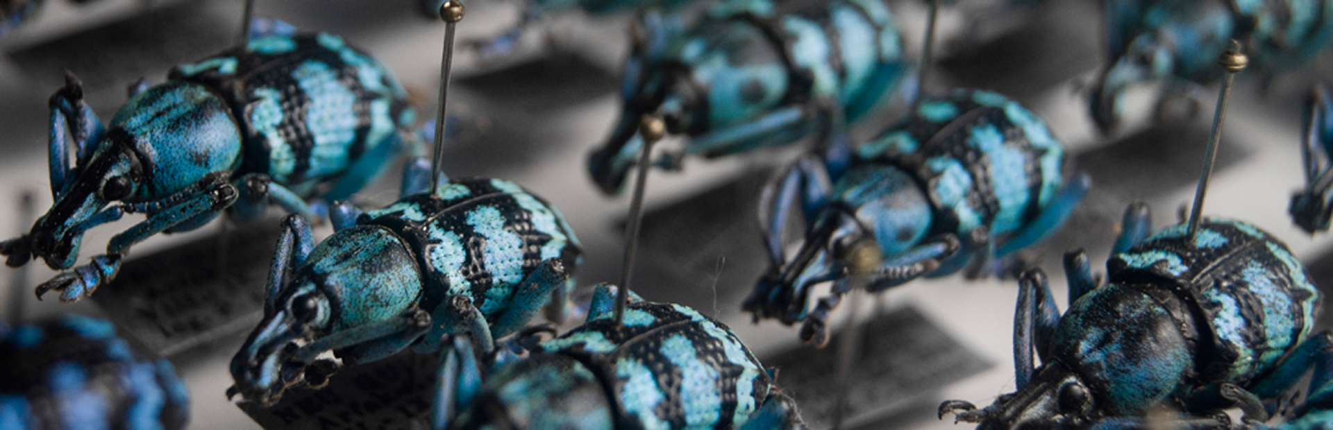
Wissenschaftskolloquium
Ansprechpartner:
Dr. Peter Warth
Tel: 0711 8936-2810
E-Mail: peter.warth(at)smns-bw.de
PD Dr. Alexander Kupfer
Tel: 0711 8936-144
E-Mail: alexander.kupfer(at)smns-bw.de
Aktuell
03.11.
Dynamic ranges, uncertain niches: A fundamental challenge in modelling species distributions under environmental change
Dr. Michael Nobis, Swiss Federal Institute for Forest, Snow and Landscape Research WSL
As usual, we will also broadcast via zoom for those who can not join in person: https://zoom.us/j/94510498226
Terminübersicht
- 06.10.
Findet leider nicht statt - 20.10.
Species delimitation 4.0: Using AI to uncover fine-scale plant phylogenomic and phenotypic variation
Dr. Kevin Karbstein, Friedrich-Schiller-Universität Jena - 03.11.
Dynamic ranges, uncertain niches: A fundamental challenge in modelling species distributions under environmental change
Dr. Michael Nobis, Swiss Federal Institute for Forest, Snow and Landscape Research WSL - 10.11.
What remains: Understanding the mechanisms of fossilisation through the lens of soft tissue preservation
Dr. Orla Bath Enright, Naturkundemuseum Stuttgart. - 17.11.
Entomonematology – Science between bugs and worms
Dr. Matthias Herrmann, Max-Planck-Institut für Biologie Tübingen - 01.12.
Titel und Referent*in folgen - 15.12.
Titel und Referent*in folgen - 19.01.
Titel und Referent*in folgen - 02.02.
Titel und Referent*in folgen